On the origin and development of the pulps and sacs of the human teeth / by John Goodsir.
- Date:
- [1839?]
Licence: Public Domain Mark
Credit: On the origin and development of the pulps and sacs of the human teeth / by John Goodsir. Source: Wellcome Collection.
Provider: This material has been provided by the Royal College of Physicians of Edinburgh. The original may be consulted at the Royal College of Physicians of Edinburgh.
12/38 (page 12)
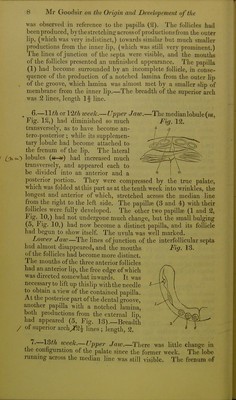
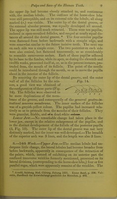
![ed in size, and was as prominent as the inner, except posteriorly where the latter still retained its posterior lobe ; but the most re- markable change which had taken place since last week Avas the complete adhesion of both lips, as in the upper jaw, with the'ex- ception of a small portion posteriorly, which still retained the pecu- liar appearance of the dental groove, and in which nothing could be seen but the smooth mucous mem- Fig. 18. branc ; {a, Fig. 18.) When the den- tal groove was torn open, as was done in the upper jaw, the lamiuje (which were highly developed) of the folli- cles, and the walls of the groove, Avere found to be rough and flocculent from adhesions, with the exception of the little depressions formerly mentioned, which still retained their original appearance. Breadth of the superior arch, 5 lines ; length, 4 lines. 10.—IQth week. — Upper Jaw.—The palate retained the appearance it had in the last subject, with the exception of the median lobule, which had become narrow in front, and broad posteriorly. The raphe of the dental groove had become firm- er, so as to give a much more defined and pennanent appear- ance to the non-adherent portion posteriorly, which was now seen to great advantage, its fine greyish mucous membrane gradually running at its edges into the white tough substance of the palate and gums. Having separated the lips of the non-adherent portion (a. Fig. 19,) a papilla, sunk in an open follicle, with Fig. 19. three or four laminse, was visible, (6.) The membrane of the palate and maxillary arch being stripped from the bone, and its sur- face of adhesion examined, lines correspond- ing with the sutures of the bones were observ- .■^/L ed; one^he median, another^the intermaxillary, and a third with the palato-maxillary. Five tooth sacs were also observed on both sides of the maxillary arch. These were divided into three groups, two in the first, or anterior, one in the second, and two in the third, or posterior. These groups Avere covered with a flocculent spongy membrane, Avhich Avas easily stripped off by the forceps, and when this was carefully done, it became evident that the sacs which Avere formerly grouped together by this mem- brane were individually isolated, and formed of a thin gray dia- ])hanous membrane, similar to the one formerly mentioned as co- vering the bottom of the dental groove, and constituting the mem- brane of the follicles. The careful detachment of the external](https://iiif.wellcomecollection.org/image/b21955451_0014.jp2/full/800%2C/0/default.jpg)