Seminiferous tubule

  • NIMR, Francis Crick Institute
  • Digital Images
  • Online

Available online

view Seminiferous tubule

Attribution-NonCommercial 4.0 International (CC BY-NC 4.0)

You can use this work for any purpose, as long as it is not primarily intended for or directed to commercial advantage or monetary compensation. You should also provide attribution to the original work, source and licence. Read more about this licence.

Credit

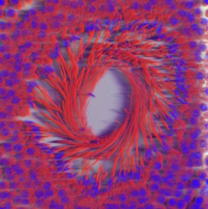
Seminiferous tubule. NIMR, Francis Crick Institute. Attribution-NonCommercial 4.0 International (CC BY-NC 4.0). Source: Wellcome Collection.

Selected images from this work

View 1 image

About this work

Description

Cross section through a seminiferous tubule showing the developing sperm with their tails pointing into the lumen of the tubule. The nuclei are stained blue with DAPI and mitochondria red with mitotracker. Sperm have a large number of mitochondria to power their swim towards the egg. The image has been volume rendered using Imaris. Wellcome Image Awards 2008.

Permanent link