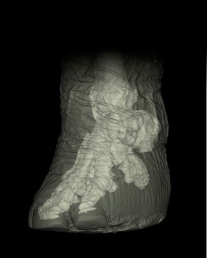
This is a reconstructed CT scan of an elephant's foot, with the exterior shown in grey and the internal skeleton in white. Elephants look flat-footed, but from the skeleton, we see that they actually walk on the tips of their toes.
The foot contains a large fat pad, and false 'sixth' toe (actually a modified sesamoid bone, at the right of the image) acts almost like a high heel to support weight.
These images are useful for researchers investigating musculoskeletal disease in elephant feet, which seems to be related to weight-bearing and age. Disease may be very common, but is hard to diagnose with x-ray partly because of the thickness of the body - x-rays are scattered and distorted, as can be seen at the base of the foot which is dense and grainy here. The images also allow analysis of modified sesamoid (false thumbs and toes) anatomy to investigate the evolution of elephants and other animals.
The width of the foot from toe to heel is around 42 centimeters.