An introduction to the study of embryology / by Alfred C. Haddon.
- Haddon, Alfred C. (Alfred Cort), 1855-1940.
- Date:
- 1887
Licence: Public Domain Mark
Credit: An introduction to the study of embryology / by Alfred C. Haddon. Source: Wellcome Collection.
Provider: This material has been provided by University of Bristol Library. The original may be consulted at University of Bristol Library.
101/374 (page 67)
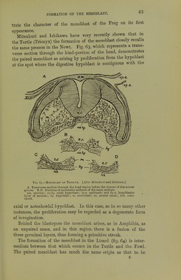
![embryo. This is formed of stellate cells, .which are at first readily distinguishable from the rounded cells of the former class; they arise from the hypoblast mainly on each side of the median line, and especially in the region in front of the primitive streak; in other words, in the embryonic region. They are continuous behind with the lateral wings of mesoblast which grow out from the primitive streak, and on their inner side are also at first continuous with the cells which form the notochord. The third portion of the mesoblast is derived partly from those cells of the lower-layer cells which do not form the permanent hypoblast, and which are scattered between that layer and the epiblast (figs. 30-34), and partly from the germinal wall, or that ridge of cells, nuclei, and yolk-granules which in the early stages FiQ. 66.—Section through the Germinal Ridge or a Fowl's Blastoderm. I-Afur Kollmann.] a. archenteron ; ep. epiblast; hy. hypoblast; m. mesoblast cells (mesamoeboids or Poreuten ) which have been derived from the pi-imitive hypoblast cells of the germinal ridge ; y. yolk; y'. yolk-spheres ingested by the primitive hypoblast. of incubation forms the marginal boundary of the lower-layer cells or primitive hypoblast (figs. 65, 66). The large primitive hypoblast cells of the germinal wall are undoubtedly nutritive in function, and ingest the underlying yolk. By cell-division they give origin to amoeboid wandering cells (fig. 66, m), which are stated by Kollmann to form the primitive vascular system, the blood, and also the connective tissue. In either case, the cells have the same morphological value since they are derived from lower- layer cells before the hypoblast proper is differentiated. While the paired mesoblast referred to above is clearly meso- thelial in character, the mesoblast which arises from the lower- layer cells and the germinal wall appears to be mesenchymatous in nature. The development of the mesoblast in .the Mole (Talpa) (fig. 67) has been shown by Heape to agree very closely with that de-](https://iiif.wellcomecollection.org/image/b21443919_0101.jp2/full/800%2C/0/default.jpg)