An introduction to the study of embryology / by Alfred C. Haddon.
- Haddon, Alfred C. (Alfred Cort), 1855-1940.
- Date:
- 1887
Licence: Public Domain Mark
Credit: An introduction to the study of embryology / by Alfred C. Haddon. Source: Wellcome Collection.
Provider: This material has been provided by University of Bristol Library. The original may be consulted at University of Bristol Library.
52/374 (page 18)
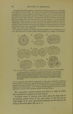
![application of staining reagents. The acliromatin is permeated by a delicate net- work or reticulum of a denser substance, the nucleoplasm or chromatin, which also forms the delicate wall of the vesicle. This network readily stains deeply, and the intersections of the fibres usually give a dotted appearance to the nucleus. When the cell is in a resting condition, the chromatin is, as a rule, concentrated either into several rounded bodies, or more frequently into a single mass, the nucleolus ; but this is usually, if not always, connected with the wall of the nucleus by delicate strands of chromatin. During the process of division in such a nucleus as that just described, the con- / A Fig. 13.—Nuclear Division. A-H. karyokinesis of a tissue-cell. A. nuclear reticulum in its ordinary state. B. preparing for division ; the contour is less defined, and tlie fibres thicker and less intricate. C. wreath-stage ; the chromatin is arranged in a complicated looping round tbe equator of the achromatin spindle. D. monaster-stage; the chromatin now appears as centripetal equatorial Vs, each of which should be re- presented as double. E. a migration of the half of each chromatin loop towai'ds opposite poles of the spindle. P. diaster-stage; the chromatin forms a star round each pole of a spindle, each aster being connected by strands of achromatin. G. daughter wreath-stage; the newly formed nuclei are passing through their retrogressive development, which is completed in the resting-stage, H. d-f. karyokinesis of an egg-cell, showing tbe smaller amount of chromatin than in the tissue-cell. The stages d. e. f. correspond to D. E. F. respectively. The polar star at the end of the spindle is composed of j^rotoplasm granules of the cell itself, and must not be mistaken for the diaster (P). The coarse lines repre- sent the chromatin, the fine lines the achromatin, and the dotted lines cell- granules {chiefly modified from Flemming]. X-Z. direct nuclear division in the cells of the embryonic integument of the European Scorpion {after Blochmann], tour becomes less defined, owing to the disappearance of its membrane ; the veiy fine close network appears looser in texture and coarser in fibre ; and a contorted looped rosette or wreath of chromatin is eventually formed (fig. 13, A-c). The peripheral loops fracture, leaving a star-like group of V-shaped bars of chromatin (aster or single star), the angles of which point towards the «entre. By this time the achromatin has been transformed into a nuclear spindle, and the chromatin wreath and single aster lie at right angles to it in its equatorial plane (c, d). Each bent chromatin bar next divides longitudinally (the division is not shown in the figure), and the loops, instead of pointing inwards, become directed, some towards one pole of the long axis of the nucleus, and some towards the other, forming a double star](https://iiif.wellcomecollection.org/image/b21443919_0052.jp2/full/800%2C/0/default.jpg)