Leishmania mexicana parasite in the promastigote stage, SEM

  • University of Oxford, Richard Wheeler
  • Digital Images
  • Online

Available online

view Leishmania mexicana parasite in the promastigote stage, SEM

Attribution 4.0 International (CC BY 4.0)

You can use this work for any purpose, including commercial uses, without restriction under copyright law. You should also provide attribution to the original work, source and licence. Read more about this licence.

Credit

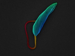
Leishmania mexicana parasite in the promastigote stage, SEM. University of Oxford, Richard Wheeler. Attribution 4.0 International (CC BY 4.0). Source: Wellcome Collection.

Selected images from this work

View 1 image

About this work

Description

Scanning electron micrograph of a Leishmania mexicana parasite. Leishmania mexicana is a unicellular eukaryotic parasite, and is one of the species of Leishmania that causes the disease Leishmaniasis. It is sometimes called the "flesh eating parasite" because of the skin lesions it can cause. These cells are in the promastigote life cycle stage; the life cycle stage normally found in the gut of sand flies. Leishmania parasites are transmitted through the bite of an infected sand fly. The parasite is approximately 8 micrometres long and 2 micrometres wide. The cell body has been pseudo-coloured green/blue, and the long flagellum has been pseudo-coloured orange/red.

Permanent link