86 results filtered with: Immunology
- Digital Images
- Online
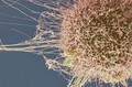
HeLa cell, immortal human epithelial cancer cell line, SEM
Anne Weston, Francis Crick Institute
- Digital Images
- Online
HeLa cell, immortal human epithelial cancer cell line, SEM
Anne Weston, Francis Crick Institute- Books
International benchmarking of US immunology research / Panel on International Benchmarking of US Immunology Research, Committee on Science, Engineering, and Public Policy, National Academy of Sciences, National Academy of Engineering, Institute of Medicine.
Date: 1999
- Digital Images
- Online
The ecology of influenza A viruses
Dolores Murcia- Audio
Virus - the unseen enemy. Pt. 3, HIV.
Date: 1999
- Digital Images
- Online
Grooved vaccination Lancet in case.

- Digital Images
- Online
Cellular architecture of normal human skin imaged by whole mount tissue microscopy. Human skin has a rich network of white blood cells (specifically dendritic cells, T cells and macrophages) which form sheaths around blood vessels. In this image, blood vessels (string-like structures stained for CD31; red), lymphatic vessels (ribbon-like structures stained for LYVE-1; blue) and dendritic cells (stained for CD11c; green) can be seen. Macrophages (stained for LYVE-1; blue) are also present. This normal cellular architecture is grossly disrupted in diseased skin (see related images). X10 magnification. Scale bar (white) represents 200 micrometres.
Dr. Xiao-nong Wang, Human Dendritic Cell Laboratory, Newcastle University
- Digital Images
- Online
HeLa cell, immortal human epithelial cancer cell line, SEM
Anne Weston, Francis Crick Institute
- Digital Images
- Online
HeLa cells, immortal human epithelial cancer cell line, SEM
Anne Weston, Francis Crick Institute
- Digital Images
- Online
Cellular architecture of normal human skin imaged by whole mount tissue microscopy. Human skin has a rich network of white blood cells (specifically dendritic cells, T cells and macrophages) which form sheaths around blood vessels. This image was taken greater than 150 micrometres beneath the junction that joins the dermal and epidermal layers of the skin (dermo-epidermal junction). At this level, dendritic cells (stained for CD11c; green) and macrophages (stained for LYVE-1; blue) form clusters around blood vessels (stained for CD31; red). This normal cellular architecture is grossly disrupted in diseased skin (see related images). Scale bar (white) represents 100 micrometres.
Dr. Xiao-nong Wang, Human Dendritic Cell Laboratory, Newcastle University
- Books
- Online
Cases of tetanus : and rabies contagiosa, or canine hydrophobia; with remarks, chiefly intended to ascertain the characteristic symptoms of the latter disease in man and certain brutes, and to point out the most effectual means of prevention. / By Caleb Hillier Parry.
Parry, Caleb Hillier, 1755-1822.Date: 1814