195 results filtered with: Development

- Digital Images
- Online
Embryonic nerves growing into the developing limb
Dr Jonathan Clarke
- Digital Images
- Online
Corrosion cast of a cow's lung
Michael Frank, Royal Veterinary College
- Digital Images
- Online
Dog hindlimb, lateral (outer) view
Michael Frank, Royal Veterinary College
- Digital Images
- Online
Skull, dugong
Michael Frank, Royal Veterinary College
- Digital Images
- Online
Lung cancer cells grown in culture, SEM
Anne Weston, Francis Crick Institute
- Digital Images
- Online
Neurons in the developing brain. The thin brown fibres are nerve cells growing out over the brain surface.
Dr Jonathan Clarke
- Digital Images
- Online
Corrosion cast of a pig's lung
Michael Frank, Royal Veterinary College
- Digital Images
- Online
HMG-CoA reductase
T. Greenhough & A. Shrive
- Digital Images
- Online
The first photo shows the physical condition of the youth of the nation as revealed by the war. The second shows what can be achieved by scientific methods of physical education and culture, and how imperative such methods are to safeguard us against physical deterioration and disease in future years.

- Digital Images
- Online
HeLa cell, immortal human epithelial cancer cell line, SEM
Anne Weston, Francis Crick Institute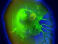
- Digital Images
- Online
6-day old chick embryo viewed under a stereo microscope, LM
Khuloud T. Al-Jamal, Serene Tay & Michael Cicirko
- Digital Images
- Online
Oligodendrocyte precursor cells (orange and yellow) in their zone of origin within the spinal cord.
Nina Callard
- Digital Images
- Online
HeLa cell, immortal human epithelial cancer cell line, SEM
Anne Weston, Francis Crick Institute
- Digital Images
- Online
Neuroepithelium, the developing brain
Prof. Bill Harris
- Digital Images
- Online
Wallaby uterus
Michael Frank, Royal Veterinary College
- Digital Images
- Online
Lung cancer cells grown in culture, SEM
Anne Weston, Francis Crick Institute
- Digital Images
- Online
2-day old zebrafish viewed under a stereo microscope, LM
Khuloud T. Al-Jamal, Serene Tay & Michael Cicirko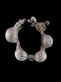
- Digital Images
- Online
Early pregnant uterus, feline
Michael Frank, Royal Veterinary College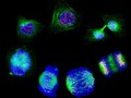
- Digital Images
- Online
Human cells showing the stages of cell division
Matthew Daniels
- Digital Images
- Online
Mouse umbilical cord
Anne Weston, Francis Crick Institute
- Digital Images
- Online
Drosophila embryo polarity - role of bicoid gene
Isabel Palacios & Daniel St Johnston
- Digital Images
- Online
Location of cyclin in 2-cell human embryo
Dr Mark Carrington,Camb.Univ.
- Digital Images
- Online
Drosophila oocyte showing actin/red and GFP-Staufen/green
Daniel St Johnston
- Digital Images
- Online
Bovine teat
Michael Frank, Royal Veterinary College
- Digital Images
- Online
Drosophila ovaries stained for actin/purple and DNA/orange.
Teresa Niccoli & Daniel St Johnston